CNSM-06 WHEN BRAIN LESIONS AREN’T WHAT THEY SEEM: THE POWER OF COMBINING MRI AND RADIOACTIVE IODINE SCINTIGRAPHY FOR THYROID CANCER IN A RESOURCE-LIMITED SETTING Article Swipe
 YOU?
·
· 2025
· Open Access
·
· DOI: https://doi.org/10.1093/noajnl/vdaf213.020
YOU?
·
· 2025
· Open Access
·
· DOI: https://doi.org/10.1093/noajnl/vdaf213.020
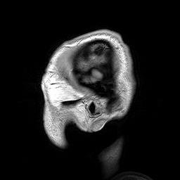
Background and Aims Accurate imaging is essential for the diagnosis, staging, and treatment planning of cancer. Magnetic Resonance Imaging (MRI) provides high-resolution anatomical detail and is preferred for evaluating intracranial lesions, while radioactive iodine (RAI) scintigraphy remains the cornerstone of functional imaging (and therapy) in well-differentiated thyroid cancer (WDTC). These modalities offer complementary insights, which are particularly valuable in resource-limited settings where hybrid imaging may be unavailable. This case report demonstrates the utility of combining MRI and RAI scintigraphy to improve diagnostic accuracy in a patient with suspected intracranial metastases from follicular thyroid cancer (FTC). Methods We retrospectively reviewed the clinical records and imaging findings (MRI and RAI scintigraphy) of a 51-year-old Nigerian woman with advanced FTC. Independent and integrated image interpretation was performed, followed by histopathological correlation. Results The patient presented with progressive right-sided exophthalmos and underwent brain MRI, which revealed a right sphenoid wing mass extending into the orbit. Initial postoperative histology suggested polymorphous low-grade adenocarcinoma, but immunohistochemistry favoured metastatic FTC. Two additional enhancing intracranial lesions were interpreted as likely benign epidermoid cysts. However, whole-body RAI scintigraphy subsequently demonstrated abnormal cranial uptake spatially congruent with the locations of these lesions, raising suspicion for metastatic disease. Surgical excision and histology confirmed the diagnosis of metastatic FTC. Conclusions Accurate staging is critical in advanced thyroid cancer, particularly when intracranial metastases are present, as these represent a contraindication to radioactive iodine therapy (RAIT) without prior surgical resection. In this case, RAI scintigraphy proved vital in reclassifying lesions initially presumed benign, thereby preventing potential complications such as cerebral edema, increased intracranial pressure, and seizures following RAIT. This case underscores the importance of combining functional and anatomical imaging to enhance diagnostic precision, especially in resource-constrained settings where hybrid imaging technologies are limited but can be effectively simulated through multimodal integration.
Related Topics


- Type
- article
- Language
- en
- Landing Page
- https://doi.org/10.1093/noajnl/vdaf213.020
- OA Status
- gold
- OpenAlex ID
- https://openalex.org/W7104440620
Raw OpenAlex JSON
- OpenAlex ID
-
https://openalex.org/W7104440620Canonical identifier for this work in OpenAlex
- DOI
-
https://doi.org/10.1093/noajnl/vdaf213.020Digital Object Identifier
- Title
-
CNSM-06 WHEN BRAIN LESIONS AREN’T WHAT THEY SEEM: THE POWER OF COMBINING MRI AND RADIOACTIVE IODINE SCINTIGRAPHY FOR THYROID CANCER IN A RESOURCE-LIMITED SETTINGWork title
- Type
-
articleOpenAlex work type
- Language
-
enPrimary language
- Publication year
-
2025Year of publication
- Publication date
-
2025-11-01Full publication date if available
- Authors
-
Peter Afolami, Augustine Adeolu, Mutiu Jimoh, Olumayowa KoladeList of authors in order
- Landing page
-
https://doi.org/10.1093/noajnl/vdaf213.020Publisher landing page
- Open access
-
YesWhether a free full text is available
- OA status
-
goldOpen access status per OpenAlex
- OA URL
-
https://doi.org/10.1093/noajnl/vdaf213.020Direct OA link when available
- Concepts
-
Medicine, Radiology, Thyroid cancer, Scintigraphy, Thyroid, Magnetic resonance imaging, Nuclear medicine, Cancer, Contraindication, Medical imaging, Context (archaeology), Neuroimaging, Anaplastic thyroid cancer, Overdiagnosis, Surgical oncologyTop concepts (fields/topics) attached by OpenAlex
- Cited by
-
0Total citation count in OpenAlex
Full payload
| id | https://openalex.org/W7104440620 |
|---|---|
| doi | https://doi.org/10.1093/noajnl/vdaf213.020 |
| ids.doi | https://doi.org/10.1093/noajnl/vdaf213.020 |
| ids.openalex | https://openalex.org/W7104440620 |
| fwci | 0.0 |
| type | article |
| title | CNSM-06 WHEN BRAIN LESIONS AREN’T WHAT THEY SEEM: THE POWER OF COMBINING MRI AND RADIOACTIVE IODINE SCINTIGRAPHY FOR THYROID CANCER IN A RESOURCE-LIMITED SETTING |
| biblio.issue | Supplement_5 |
| biblio.volume | 7 |
| biblio.last_page | |
| biblio.first_page | |
| topics[0].id | https://openalex.org/T10329 |
| topics[0].field.id | https://openalex.org/fields/27 |
| topics[0].field.display_name | Medicine |
| topics[0].score | 0.8011462092399597 |
| topics[0].domain.id | https://openalex.org/domains/4 |
| topics[0].domain.display_name | Health Sciences |
| topics[0].subfield.id | https://openalex.org/subfields/2712 |
| topics[0].subfield.display_name | Endocrinology, Diabetes and Metabolism |
| topics[0].display_name | Thyroid Cancer Diagnosis and Treatment |
| topics[1].id | https://openalex.org/T11538 |
| topics[1].field.id | https://openalex.org/fields/27 |
| topics[1].field.display_name | Medicine |
| topics[1].score | 0.061611078679561615 |
| topics[1].domain.id | https://openalex.org/domains/4 |
| topics[1].domain.display_name | Health Sciences |
| topics[1].subfield.id | https://openalex.org/subfields/2734 |
| topics[1].subfield.display_name | Pathology and Forensic Medicine |
| topics[1].display_name | Ophthalmology and Eye Disorders |
| topics[2].id | https://openalex.org/T11328 |
| topics[2].field.id | https://openalex.org/fields/27 |
| topics[2].field.display_name | Medicine |
| topics[2].score | 0.02718915231525898 |
| topics[2].domain.id | https://openalex.org/domains/4 |
| topics[2].domain.display_name | Health Sciences |
| topics[2].subfield.id | https://openalex.org/subfields/2731 |
| topics[2].subfield.display_name | Ophthalmology |
| topics[2].display_name | Ocular Oncology and Treatments |
| is_xpac | False |
| apc_list.value | 1622 |
| apc_list.currency | GBP |
| apc_list.value_usd | 1989 |
| apc_paid.value | 1622 |
| apc_paid.currency | GBP |
| apc_paid.value_usd | 1989 |
| concepts[0].id | https://openalex.org/C71924100 |
| concepts[0].level | 0 |
| concepts[0].score | 0.9355599880218506 |
| concepts[0].wikidata | https://www.wikidata.org/wiki/Q11190 |
| concepts[0].display_name | Medicine |
| concepts[1].id | https://openalex.org/C126838900 |
| concepts[1].level | 1 |
| concepts[1].score | 0.7047756910324097 |
| concepts[1].wikidata | https://www.wikidata.org/wiki/Q77604 |
| concepts[1].display_name | Radiology |
| concepts[2].id | https://openalex.org/C2779761222 |
| concepts[2].level | 3 |
| concepts[2].score | 0.6311274170875549 |
| concepts[2].wikidata | https://www.wikidata.org/wiki/Q826522 |
| concepts[2].display_name | Thyroid cancer |
| concepts[3].id | https://openalex.org/C2779902710 |
| concepts[3].level | 2 |
| concepts[3].score | 0.6186431646347046 |
| concepts[3].wikidata | https://www.wikidata.org/wiki/Q1130465 |
| concepts[3].display_name | Scintigraphy |
| concepts[4].id | https://openalex.org/C526584372 |
| concepts[4].level | 2 |
| concepts[4].score | 0.5581462979316711 |
| concepts[4].wikidata | https://www.wikidata.org/wiki/Q16399 |
| concepts[4].display_name | Thyroid |
| concepts[5].id | https://openalex.org/C143409427 |
| concepts[5].level | 2 |
| concepts[5].score | 0.542091429233551 |
| concepts[5].wikidata | https://www.wikidata.org/wiki/Q161238 |
| concepts[5].display_name | Magnetic resonance imaging |
| concepts[6].id | https://openalex.org/C2989005 |
| concepts[6].level | 1 |
| concepts[6].score | 0.36132657527923584 |
| concepts[6].wikidata | https://www.wikidata.org/wiki/Q214963 |
| concepts[6].display_name | Nuclear medicine |
| concepts[7].id | https://openalex.org/C121608353 |
| concepts[7].level | 2 |
| concepts[7].score | 0.32492417097091675 |
| concepts[7].wikidata | https://www.wikidata.org/wiki/Q12078 |
| concepts[7].display_name | Cancer |
| concepts[8].id | https://openalex.org/C2776494729 |
| concepts[8].level | 3 |
| concepts[8].score | 0.3122521936893463 |
| concepts[8].wikidata | https://www.wikidata.org/wiki/Q1848900 |
| concepts[8].display_name | Contraindication |
| concepts[9].id | https://openalex.org/C31601959 |
| concepts[9].level | 2 |
| concepts[9].score | 0.30041244626045227 |
| concepts[9].wikidata | https://www.wikidata.org/wiki/Q931309 |
| concepts[9].display_name | Medical imaging |
| concepts[10].id | https://openalex.org/C2779343474 |
| concepts[10].level | 2 |
| concepts[10].score | 0.27966105937957764 |
| concepts[10].wikidata | https://www.wikidata.org/wiki/Q3109175 |
| concepts[10].display_name | Context (archaeology) |
| concepts[11].id | https://openalex.org/C58693492 |
| concepts[11].level | 2 |
| concepts[11].score | 0.279354989528656 |
| concepts[11].wikidata | https://www.wikidata.org/wiki/Q551875 |
| concepts[11].display_name | Neuroimaging |
| concepts[12].id | https://openalex.org/C2780256643 |
| concepts[12].level | 4 |
| concepts[12].score | 0.2712458074092865 |
| concepts[12].wikidata | https://www.wikidata.org/wiki/Q3658382 |
| concepts[12].display_name | Anaplastic thyroid cancer |
| concepts[13].id | https://openalex.org/C2779377019 |
| concepts[13].level | 2 |
| concepts[13].score | 0.26466602087020874 |
| concepts[13].wikidata | https://www.wikidata.org/wiki/Q956131 |
| concepts[13].display_name | Overdiagnosis |
| concepts[14].id | https://openalex.org/C2780140570 |
| concepts[14].level | 2 |
| concepts[14].score | 0.256877601146698 |
| concepts[14].wikidata | https://www.wikidata.org/wiki/Q3545481 |
| concepts[14].display_name | Surgical oncology |
| keywords[0].id | https://openalex.org/keywords/thyroid-cancer |
| keywords[0].score | 0.6311274170875549 |
| keywords[0].display_name | Thyroid cancer |
| keywords[1].id | https://openalex.org/keywords/scintigraphy |
| keywords[1].score | 0.6186431646347046 |
| keywords[1].display_name | Scintigraphy |
| keywords[2].id | https://openalex.org/keywords/thyroid |
| keywords[2].score | 0.5581462979316711 |
| keywords[2].display_name | Thyroid |
| keywords[3].id | https://openalex.org/keywords/magnetic-resonance-imaging |
| keywords[3].score | 0.542091429233551 |
| keywords[3].display_name | Magnetic resonance imaging |
| keywords[4].id | https://openalex.org/keywords/cancer |
| keywords[4].score | 0.32492417097091675 |
| keywords[4].display_name | Cancer |
| keywords[5].id | https://openalex.org/keywords/contraindication |
| keywords[5].score | 0.3122521936893463 |
| keywords[5].display_name | Contraindication |
| language | en |
| locations[0].id | doi:10.1093/noajnl/vdaf213.020 |
| locations[0].is_oa | True |
| locations[0].source.id | https://openalex.org/S4210237914 |
| locations[0].source.issn | 2632-2498 |
| locations[0].source.type | journal |
| locations[0].source.is_oa | True |
| locations[0].source.issn_l | 2632-2498 |
| locations[0].source.is_core | True |
| locations[0].source.is_in_doaj | True |
| locations[0].source.display_name | Neuro-Oncology Advances |
| locations[0].source.host_organization | https://openalex.org/P4310311648 |
| locations[0].source.host_organization_name | Oxford University Press |
| locations[0].source.host_organization_lineage | https://openalex.org/P4310311648 |
| locations[0].license | cc-by-nc |
| locations[0].pdf_url | |
| locations[0].version | publishedVersion |
| locations[0].raw_type | journal-article |
| locations[0].license_id | https://openalex.org/licenses/cc-by-nc |
| locations[0].is_accepted | True |
| locations[0].is_published | True |
| locations[0].raw_source_name | Neuro-Oncology Advances |
| locations[0].landing_page_url | https://doi.org/10.1093/noajnl/vdaf213.020 |
| indexed_in | crossref, doaj |
| authorships[0].author.id | https://openalex.org/A3216574535 |
| authorships[0].author.orcid | |
| authorships[0].author.display_name | Peter Afolami |
| authorships[0].affiliations[0].raw_affiliation_string | University College Hospital , Ibadan, |
| authorships[0].author_position | first |
| authorships[0].raw_author_name | Peter Afolami |
| authorships[0].is_corresponding | True |
| authorships[0].raw_affiliation_strings | University College Hospital , Ibadan, |
| authorships[1].author.id | https://openalex.org/A4222928752 |
| authorships[1].author.orcid | |
| authorships[1].author.display_name | Augustine Adeolu |
| authorships[1].affiliations[0].raw_affiliation_string | University College Hospital , Ibadan, |
| authorships[1].affiliations[1].raw_affiliation_string | University of Ibadan , Ibadan, |
| authorships[1].author_position | middle |
| authorships[1].raw_author_name | Augustine Adeolu |
| authorships[1].is_corresponding | False |
| authorships[1].raw_affiliation_strings | University College Hospital , Ibadan,, University of Ibadan , Ibadan, |
| authorships[2].author.id | https://openalex.org/A2650150491 |
| authorships[2].author.orcid | https://orcid.org/0000-0002-6383-3961 |
| authorships[2].author.display_name | Mutiu Jimoh |
| authorships[2].affiliations[0].raw_affiliation_string | University College Hospital , Ibadan, |
| authorships[2].affiliations[1].raw_affiliation_string | University of Ibadan , Ibadan, |
| authorships[2].author_position | middle |
| authorships[2].raw_author_name | Mutiu Jimoh |
| authorships[2].is_corresponding | False |
| authorships[2].raw_affiliation_strings | University College Hospital , Ibadan,, University of Ibadan , Ibadan, |
| authorships[3].author.id | https://openalex.org/A4380454454 |
| authorships[3].author.orcid | https://orcid.org/0000-0001-8551-1114 |
| authorships[3].author.display_name | Olumayowa Kolade |
| authorships[3].affiliations[0].raw_affiliation_string | University College Hospital , Ibadan, |
| authorships[3].author_position | last |
| authorships[3].raw_author_name | Olumayowa Kolade |
| authorships[3].is_corresponding | False |
| authorships[3].raw_affiliation_strings | University College Hospital , Ibadan, |
| has_content.pdf | False |
| has_content.grobid_xml | False |
| is_paratext | False |
| open_access.is_oa | True |
| open_access.oa_url | https://doi.org/10.1093/noajnl/vdaf213.020 |
| open_access.oa_status | gold |
| open_access.any_repository_has_fulltext | False |
| created_date | 2025-11-09T00:00:00 |
| display_name | CNSM-06 WHEN BRAIN LESIONS AREN’T WHAT THEY SEEM: THE POWER OF COMBINING MRI AND RADIOACTIVE IODINE SCINTIGRAPHY FOR THYROID CANCER IN A RESOURCE-LIMITED SETTING |
| has_fulltext | False |
| is_retracted | False |
| updated_date | 2025-11-09T23:12:50.468023 |
| primary_topic.id | https://openalex.org/T10329 |
| primary_topic.field.id | https://openalex.org/fields/27 |
| primary_topic.field.display_name | Medicine |
| primary_topic.score | 0.8011462092399597 |
| primary_topic.domain.id | https://openalex.org/domains/4 |
| primary_topic.domain.display_name | Health Sciences |
| primary_topic.subfield.id | https://openalex.org/subfields/2712 |
| primary_topic.subfield.display_name | Endocrinology, Diabetes and Metabolism |
| primary_topic.display_name | Thyroid Cancer Diagnosis and Treatment |
| cited_by_count | 0 |
| locations_count | 1 |
| best_oa_location.id | doi:10.1093/noajnl/vdaf213.020 |
| best_oa_location.is_oa | True |
| best_oa_location.source.id | https://openalex.org/S4210237914 |
| best_oa_location.source.issn | 2632-2498 |
| best_oa_location.source.type | journal |
| best_oa_location.source.is_oa | True |
| best_oa_location.source.issn_l | 2632-2498 |
| best_oa_location.source.is_core | True |
| best_oa_location.source.is_in_doaj | True |
| best_oa_location.source.display_name | Neuro-Oncology Advances |
| best_oa_location.source.host_organization | https://openalex.org/P4310311648 |
| best_oa_location.source.host_organization_name | Oxford University Press |
| best_oa_location.source.host_organization_lineage | https://openalex.org/P4310311648 |
| best_oa_location.license | cc-by-nc |
| best_oa_location.pdf_url | |
| best_oa_location.version | publishedVersion |
| best_oa_location.raw_type | journal-article |
| best_oa_location.license_id | https://openalex.org/licenses/cc-by-nc |
| best_oa_location.is_accepted | True |
| best_oa_location.is_published | True |
| best_oa_location.raw_source_name | Neuro-Oncology Advances |
| best_oa_location.landing_page_url | https://doi.org/10.1093/noajnl/vdaf213.020 |
| primary_location.id | doi:10.1093/noajnl/vdaf213.020 |
| primary_location.is_oa | True |
| primary_location.source.id | https://openalex.org/S4210237914 |
| primary_location.source.issn | 2632-2498 |
| primary_location.source.type | journal |
| primary_location.source.is_oa | True |
| primary_location.source.issn_l | 2632-2498 |
| primary_location.source.is_core | True |
| primary_location.source.is_in_doaj | True |
| primary_location.source.display_name | Neuro-Oncology Advances |
| primary_location.source.host_organization | https://openalex.org/P4310311648 |
| primary_location.source.host_organization_name | Oxford University Press |
| primary_location.source.host_organization_lineage | https://openalex.org/P4310311648 |
| primary_location.license | cc-by-nc |
| primary_location.pdf_url | |
| primary_location.version | publishedVersion |
| primary_location.raw_type | journal-article |
| primary_location.license_id | https://openalex.org/licenses/cc-by-nc |
| primary_location.is_accepted | True |
| primary_location.is_published | True |
| primary_location.raw_source_name | Neuro-Oncology Advances |
| primary_location.landing_page_url | https://doi.org/10.1093/noajnl/vdaf213.020 |
| publication_date | 2025-11-01 |
| publication_year | 2025 |
| referenced_works_count | 0 |
| abstract_inverted_index.a | 85, 111, 143, 226 |
| abstract_inverted_index.In | 237 |
| abstract_inverted_index.We | 97 |
| abstract_inverted_index.as | 171, 223, 255 |
| abstract_inverted_index.be | 66, 292 |
| abstract_inverted_index.by | 126 |
| abstract_inverted_index.in | 45, 59, 84, 213, 244, 281 |
| abstract_inverted_index.is | 6, 26, 211 |
| abstract_inverted_index.of | 15, 40, 74, 110, 190, 205, 270 |
| abstract_inverted_index.to | 80, 228, 276 |
| abstract_inverted_index.MRI | 76 |
| abstract_inverted_index.RAI | 78, 108, 178, 240 |
| abstract_inverted_index.The | 130 |
| abstract_inverted_index.Two | 164 |
| abstract_inverted_index.and | 2, 12, 25, 77, 103, 107, 119, 137, 200, 261, 273 |
| abstract_inverted_index.are | 56, 221, 288 |
| abstract_inverted_index.but | 159, 290 |
| abstract_inverted_index.can | 291 |
| abstract_inverted_index.for | 8, 28, 195 |
| abstract_inverted_index.may | 65 |
| abstract_inverted_index.the | 9, 38, 72, 100, 150, 188, 203, 268 |
| abstract_inverted_index.was | 123 |
| abstract_inverted_index.(MRI | 106 |
| abstract_inverted_index.(and | 43 |
| abstract_inverted_index.Aims | 3 |
| abstract_inverted_index.FTC. | 117, 163, 207 |
| abstract_inverted_index.MRI, | 140 |
| abstract_inverted_index.This | 68, 265 |
| abstract_inverted_index.case | 69, 266 |
| abstract_inverted_index.from | 91 |
| abstract_inverted_index.into | 149 |
| abstract_inverted_index.mass | 147 |
| abstract_inverted_index.such | 254 |
| abstract_inverted_index.this | 238 |
| abstract_inverted_index.were | 169 |
| abstract_inverted_index.when | 218 |
| abstract_inverted_index.wing | 146 |
| abstract_inverted_index.with | 87, 115, 133, 187 |
| abstract_inverted_index.(MRI) | 20 |
| abstract_inverted_index.(RAI) | 35 |
| abstract_inverted_index.RAIT. | 264 |
| abstract_inverted_index.These | 50 |
| abstract_inverted_index.brain | 139 |
| abstract_inverted_index.case, | 239 |
| abstract_inverted_index.image | 121 |
| abstract_inverted_index.offer | 52 |
| abstract_inverted_index.prior | 234 |
| abstract_inverted_index.right | 144 |
| abstract_inverted_index.these | 191, 224 |
| abstract_inverted_index.vital | 243 |
| abstract_inverted_index.where | 62, 284 |
| abstract_inverted_index.which | 55, 141 |
| abstract_inverted_index.while | 32 |
| abstract_inverted_index.woman | 114 |
| abstract_inverted_index.(FTC). | 95 |
| abstract_inverted_index.(RAIT) | 232 |
| abstract_inverted_index.benign | 173 |
| abstract_inverted_index.cancer | 48, 94 |
| abstract_inverted_index.cysts. | 175 |
| abstract_inverted_index.detail | 24 |
| abstract_inverted_index.edema, | 257 |
| abstract_inverted_index.hybrid | 63, 285 |
| abstract_inverted_index.iodine | 34, 230 |
| abstract_inverted_index.likely | 172 |
| abstract_inverted_index.orbit. | 151 |
| abstract_inverted_index.proved | 242 |
| abstract_inverted_index.report | 70 |
| abstract_inverted_index.uptake | 184 |
| abstract_inverted_index.(WDTC). | 49 |
| abstract_inverted_index.Imaging | 19 |
| abstract_inverted_index.Initial | 152 |
| abstract_inverted_index.Methods | 96 |
| abstract_inverted_index.Results | 129 |
| abstract_inverted_index.benign, | 249 |
| abstract_inverted_index.cancer, | 216 |
| abstract_inverted_index.cancer. | 16 |
| abstract_inverted_index.cranial | 183 |
| abstract_inverted_index.enhance | 277 |
| abstract_inverted_index.imaging | 5, 42, 64, 104, 275, 286 |
| abstract_inverted_index.improve | 81 |
| abstract_inverted_index.lesions | 168, 246 |
| abstract_inverted_index.limited | 289 |
| abstract_inverted_index.patient | 86, 131 |
| abstract_inverted_index.raising | 193 |
| abstract_inverted_index.records | 102 |
| abstract_inverted_index.remains | 37 |
| abstract_inverted_index.staging | 210 |
| abstract_inverted_index.therapy | 231 |
| abstract_inverted_index.thereby | 250 |
| abstract_inverted_index.through | 295 |
| abstract_inverted_index.thyroid | 47, 93, 215 |
| abstract_inverted_index.utility | 73 |
| abstract_inverted_index.without | 233 |
| abstract_inverted_index.Abstract | 0 |
| abstract_inverted_index.Accurate | 4, 209 |
| abstract_inverted_index.However, | 176 |
| abstract_inverted_index.Magnetic | 17 |
| abstract_inverted_index.Nigerian | 113 |
| abstract_inverted_index.Surgical | 198 |
| abstract_inverted_index.abnormal | 182 |
| abstract_inverted_index.accuracy | 83 |
| abstract_inverted_index.advanced | 116, 214 |
| abstract_inverted_index.cerebral | 256 |
| abstract_inverted_index.clinical | 101 |
| abstract_inverted_index.critical | 212 |
| abstract_inverted_index.disease. | 197 |
| abstract_inverted_index.excision | 199 |
| abstract_inverted_index.favoured | 161 |
| abstract_inverted_index.findings | 105 |
| abstract_inverted_index.followed | 125 |
| abstract_inverted_index.lesions, | 31, 192 |
| abstract_inverted_index.planning | 14 |
| abstract_inverted_index.present, | 222 |
| abstract_inverted_index.presumed | 248 |
| abstract_inverted_index.provides | 21 |
| abstract_inverted_index.revealed | 142 |
| abstract_inverted_index.reviewed | 99 |
| abstract_inverted_index.seizures | 262 |
| abstract_inverted_index.settings | 61, 283 |
| abstract_inverted_index.sphenoid | 145 |
| abstract_inverted_index.staging, | 11 |
| abstract_inverted_index.surgical | 235 |
| abstract_inverted_index.therapy) | 44 |
| abstract_inverted_index.valuable | 58 |
| abstract_inverted_index.Resonance | 18 |
| abstract_inverted_index.combining | 75, 271 |
| abstract_inverted_index.confirmed | 202 |
| abstract_inverted_index.congruent | 186 |
| abstract_inverted_index.diagnosis | 204 |
| abstract_inverted_index.enhancing | 166 |
| abstract_inverted_index.essential | 7 |
| abstract_inverted_index.extending | 148 |
| abstract_inverted_index.following | 263 |
| abstract_inverted_index.histology | 154, 201 |
| abstract_inverted_index.increased | 258 |
| abstract_inverted_index.initially | 247 |
| abstract_inverted_index.insights, | 54 |
| abstract_inverted_index.locations | 189 |
| abstract_inverted_index.low-grade | 157 |
| abstract_inverted_index.potential | 252 |
| abstract_inverted_index.preferred | 27 |
| abstract_inverted_index.presented | 132 |
| abstract_inverted_index.pressure, | 260 |
| abstract_inverted_index.represent | 225 |
| abstract_inverted_index.simulated | 294 |
| abstract_inverted_index.spatially | 185 |
| abstract_inverted_index.suggested | 155 |
| abstract_inverted_index.suspected | 88 |
| abstract_inverted_index.suspicion | 194 |
| abstract_inverted_index.treatment | 13 |
| abstract_inverted_index.underwent | 138 |
| abstract_inverted_index.Background | 1 |
| abstract_inverted_index.additional | 165 |
| abstract_inverted_index.anatomical | 23, 274 |
| abstract_inverted_index.diagnosis, | 10 |
| abstract_inverted_index.diagnostic | 82, 278 |
| abstract_inverted_index.epidermoid | 174 |
| abstract_inverted_index.especially | 280 |
| abstract_inverted_index.evaluating | 29 |
| abstract_inverted_index.follicular | 92 |
| abstract_inverted_index.functional | 41, 272 |
| abstract_inverted_index.importance | 269 |
| abstract_inverted_index.integrated | 120 |
| abstract_inverted_index.metastases | 90, 220 |
| abstract_inverted_index.metastatic | 162, 196, 206 |
| abstract_inverted_index.modalities | 51 |
| abstract_inverted_index.multimodal | 296 |
| abstract_inverted_index.performed, | 124 |
| abstract_inverted_index.precision, | 279 |
| abstract_inverted_index.preventing | 251 |
| abstract_inverted_index.resection. | 236 |
| abstract_inverted_index.whole-body | 177 |
| abstract_inverted_index.51-year-old | 112 |
| abstract_inverted_index.Conclusions | 208 |
| abstract_inverted_index.Independent | 118 |
| abstract_inverted_index.cornerstone | 39 |
| abstract_inverted_index.effectively | 293 |
| abstract_inverted_index.interpreted | 170 |
| abstract_inverted_index.progressive | 134 |
| abstract_inverted_index.radioactive | 33, 229 |
| abstract_inverted_index.right-sided | 135 |
| abstract_inverted_index.underscores | 267 |
| abstract_inverted_index.correlation. | 128 |
| abstract_inverted_index.demonstrated | 181 |
| abstract_inverted_index.demonstrates | 71 |
| abstract_inverted_index.exophthalmos | 136 |
| abstract_inverted_index.integration. | 297 |
| abstract_inverted_index.intracranial | 30, 89, 167, 219, 259 |
| abstract_inverted_index.particularly | 57, 217 |
| abstract_inverted_index.polymorphous | 156 |
| abstract_inverted_index.scintigraphy | 36, 79, 179, 241 |
| abstract_inverted_index.subsequently | 180 |
| abstract_inverted_index.technologies | 287 |
| abstract_inverted_index.unavailable. | 67 |
| abstract_inverted_index.complementary | 53 |
| abstract_inverted_index.complications | 253 |
| abstract_inverted_index.postoperative | 153 |
| abstract_inverted_index.reclassifying | 245 |
| abstract_inverted_index.scintigraphy) | 109 |
| abstract_inverted_index.interpretation | 122 |
| abstract_inverted_index.adenocarcinoma, | 158 |
| abstract_inverted_index.high-resolution | 22 |
| abstract_inverted_index.retrospectively | 98 |
| abstract_inverted_index.contraindication | 227 |
| abstract_inverted_index.resource-limited | 60 |
| abstract_inverted_index.histopathological | 127 |
| abstract_inverted_index.well-differentiated | 46 |
| abstract_inverted_index.immunohistochemistry | 160 |
| abstract_inverted_index.resource-constrained | 282 |
| cited_by_percentile_year | |
| countries_distinct_count | 0 |
| institutions_distinct_count | 4 |
| citation_normalized_percentile.value | 0.79168311 |
| citation_normalized_percentile.is_in_top_1_percent | False |
| citation_normalized_percentile.is_in_top_10_percent | False |