Commentary: Can machine be taught to detect retinoblastoma? Article Swipe
 YOU?
·
· 2023
· Open Access
·
· DOI: https://doi.org/10.4103/ijo.ijo_2283_22
· OA: W4318916939
YOU?
·
· 2023
· Open Access
·
· DOI: https://doi.org/10.4103/ijo.ijo_2283_22
· OA: W4318916939
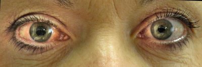
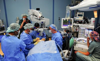
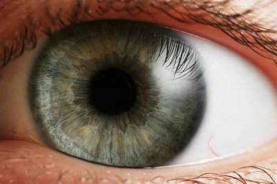
Retinoblastoma (RB) is the most common intraocular malignant tumor in children and leukocoria or white pupillary reflex is the most common and earliest clinical sign. Detection of leukocoria is dependent on the tumor size, location, ambient lighting condition, status of the pupil, etc. While tumors at the posterior pole are likely to present with early leukocoria, peripheral tumors can present as leukocoria only when it has grown to a sizeable extent. Hence, in the current treatment era of RB while we are talking not only about eye salvage but vision salvage, early detection with leukocoria as the screening strategy might not be early enough. Current screening strategies for early RB detection are mostly based on red reflex testing to detect a white glow using either an ophthalmoscope, retinoscope, camera, mobile apps, etc. These strategies can screen out children who need further evaluation and have variable sensitivity and specificity. Applicability of artificial intelligence (AI) and machine learning (ML) in retinoblastoma can be either for screening or for arriving at a diagnosis.[1,2] Screening for retinal diseases using retinal images and using AI and ML for diagnosis has been used for glaucoma, diabetic retinopathy (DR) as well as retinopathy of prematurity (ROP).[3-6] AI-based screening tools can also have a potential application for follow-up of treated RB patients and can triage the patients who need to travel to a tertiary care treating facility. However, unlike other retinal diseases like glaucoma, DR, ROP, etc., where the disease presentation and progression has a predictable course, the clinical appearance, location, and features of RB are variable and follows a more unpredictable course. Any AI-based detection algorithm needs to be robust enough to consider these factors to have improved prediction accuracy. Image quality is an important factor for any machine learning algorithm. While the application of current AI-based diagnostic algorithms has been developed for diseases located in the posterior pole of the retina where good quality images can be captured easily, retinal location of RB varies with the age at presentation and distortion while capturing peripheral retinal images might hamper the AI-based diagnosis. This aspect needs to be addressed for any AI-based diagnosis and grouping of RB. Also, in the absence of a high-risk population to screen, any screening and early diagnosis tool for RB should be portable, easy to use, should be integrable with other pediatric eye screening activities like under-five eye screening at the community, screening of the newborns in neonatal units by pediatricians, etc. The currently available wide-field retinal imaging systems required for this purpose are mostly not portable, highly expensive, and need trained personal as well as cooperation from the child for obtaining a good quality image. RB images used for training any AI model are likely to be obtained from children under anesthesia and might not be replicative of the images obtained in the field where the cooperation level of the child might affect the image quality. Unless, these aspects are taken care of, implementing and using AI for RB screening will have limited application. The rarity and low prevalence of RB in the general population make a mass screening strategy using AI and retinal imaging impractical. Piggybacking it with other neonatal and pediatric eye screening program will likely miss out on screening older children (>5 years) who are more likely to have a peripheral retinal tumor, with leukocoria in later stages. Hence, simultaneous work of designing a low-cost wide-field retinal imaging system capable of capturing good quality standardized retinal images is a necessity for any AI-based screening and diagnosis of RB to have meaningful utility. The current RB screening strategy uses high-risk and genetics-based screening. Siblings of RB children are mostly screened as per this strategy. For this cohort, AI-based screening and diagnosis can possibly obviate the need for an examination under anesthesia for many of them. Diagnosis of RB based on AI platform though technically feasible can have associated challenges. Gold standard for diagnosis of RB is the clinical evaluation by an expert preferably an examination under anesthesia with a detailed anterior and posterior segment evaluation. Hence, image-based diagnosis should consider not only interpretation of retinal images but anterior segment findings like the presence of anterior chamber seeding, neovascularization, etc. Tumors can also be situated at the periphery, which might not be detectable on conventional wide filed fundus imaging unless indentation is done and hence might have false-negative interpretation. The need for medical accuracy for diagnosis of RB is high considering the life-threatening nature of the condition, the cohort of patients who might not be able to articulate their problem, the psychosocial implications on the family, and consequently false negative results can have devastating consequences. Moreover, many simulating lesions with overlapping clinical features can confound and affect the prediction accuracy of the machine unless extensive training of the model is done to exclude these simulators also. Finally, all early detection and screening strategies are likely to be beneficial for the population and the regions with a high disease burden. RB predominantly is a disease of the low socioeconomic strata and half of the world's RB children reside in the low- and middle-income countries like Latin America, Africa, and India where majority of the children unfortunately still present with the extraocular disease with high mortality.[7] Retinal image-based diagnosis is not applicable in these scenarios. Lack of awareness about the disease, especially at the parental and community level is a major reason for not seeking early eye care consult by these families. Hence, the strategy for improving RB outcome in these countries need to focus on awareness initiatives and mobilize resources in this direction and encourage and educate families on the early signs as well as on the need for routine eye screening of children. Unless that is done, the availability of a robust machine learning system will not have enough application to reach out to each and every child at the doorstep and clinch the diagnosis at a stage where we talk of vision salvage and not only survival in RB.