Comparison of Optical Coherence Tomography With Fundus Photographs, Fluorescein Angiography, and Histopathologic Analysis in Assessing Coats Disease Article Swipe
Related Concepts
Sally S. Ong
,
Thomas J. Cummings
,
Lejla Vajzovic
,
Prithvi Mruthyunjaya
,
Cynthia A. Toth
·
 YOU?
·
· 2018
· Open Access
·
· DOI: https://doi.org/10.1001/jamaophthalmol.2018.5654
· OA: W2903300500
YOU?
·
· 2018
· Open Access
·
· DOI: https://doi.org/10.1001/jamaophthalmol.2018.5654
· OA: W2903300500
YOU?
·
· 2018
· Open Access
·
· DOI: https://doi.org/10.1001/jamaophthalmol.2018.5654
· OA: W2903300500
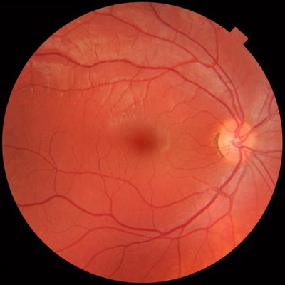
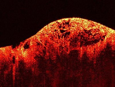
Optical coherence tomography may show the transient and permanent effects of Coats disease on the retina. These results suggest that exudates and fluid in the macular subretinal space appear later in the disease and may result in fibrosis formation. Further studies are needed to confirm if early treatment could prevent vision-threatening macular fibrosis.
Related Topics


Finding more related topics…